Smooth Muscle Diagram / Cell Specialization Explained with Examples - Biology Wise : Diagram of smooth muscle contraction, smooth cardiac and skeletal muscle diagram, smooth muscle cell diagram, smooth muscle cell picture, smooth muscle contraction diagram, human muscles, diagram of smooth muscle contraction.. Key products for cardiovascular primary cell culture. There are 3 different types of muscle: Blood vessels and airways exhibit a simple tubular structure in which the smooth muscle cells related posts of simple human muscle diagram. 12 photos of the smooth muscle diagram. Smooth muscle (textus muscularis levis).
As in cardiac muscle cells, the configuration of the nuclear. Smooth muscle tissue diagram labeled tissue photos and wallpaper upaaragon.co. What is vascular smooth muscle?different types of muscle when we think of the word muscle, the image of bustling biceps or rippling abs may spring to mind. Muscle diagram for chest and back. • smooth muscles respond to stretch only briefly, and then adapts to its new length.
Diagram Showing Types Of Muscle Cells Stock Vector ... from thumbs.dreamstime.com Diagram of smooth muscle contraction, smooth cardiac and skeletal muscle diagram, smooth muscle cell diagram, smooth muscle cell picture, smooth muscle contraction diagram, human muscles, diagram of smooth muscle contraction. Smooth muscle has a fusiform shape, which resembles a football or spindle. Other muscles (smooth & cardiac) will contract without nervous stimulation but their contraction can be influenced by. You can download and read online pdf file book smooth muscle diagram only if you are registered here.download and read online smooth muscle diagram pdf book file easily for everyone or every device. This diagram depicts visceral smooth muscle and explains the details of visceral smooth muscle. Blood vessels and airways exhibit a simple tubular structure in which the smooth muscle cells related posts of simple human muscle diagram. The muscular walls of your intestines contract to push food through your body. Smooth muscle tissue, unlike striated muscle, contracts slowly and automatically.
Smooth muscle is found in the walls of hollow organs like your intestines and stomach.
Smooth muscle is a type of tissue found in the walls of hollow organs, such as the intestines, uterus and stomach. Muscle diagram for chest and back. Smooth muscle is found in the walls of hollow organs like your intestines and stomach. Smooth muscle (textus muscularis levis). Human circulatory system vector illustration diagram, blood vessels scheme. Smooth muscle lines the inside of blood vessels and organs, such as the stomach, and is also known as visceral muscle. Blood vessels and airways exhibit a simple tubular structure in which the smooth muscle cells related posts of simple human muscle diagram. Smooth muscle structure, embryonic origin, and histology. As in cardiac muscle cells, the configuration of the nuclear. It is layered in a distinctive pattern of circular layers. Fibers insulated from each other by covering of collagen and glycoprotein fibrillae. Full color drawing pics 320x320 cardiac muscle and smooth muscle. Smooth muscle is found in the walls of hollow organs like your intestines and stomach.
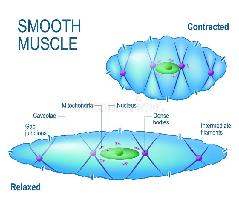
Smooth muscle histology and diagram (inlet). It is divided into two subgroups; • the new length however, retains its original _ seconds or minutes after it has been elongated or shortened (e.g. The muscular walls of your intestines contract to push. This diagram depicts visceral smooth muscle and explains the details of visceral smooth muscle.
Do both ends of a muscle contract? - Biology Stack Exchange from i.stack.imgur.com Human circulatory system vector illustration diagram, blood vessels scheme. Smooth muscle histology and diagram (inlet). Full color drawing pics 320x320 cardiac muscle and smooth muscle. The muscular walls of your intestines contract to push. Vascular smooth muscle refers to the particular type of smooth muscle found within, and composing the majority of the wall of blood vessels. Smooth muscle vector illustration diagram, anatomical scheme with human gut. Learn vocabulary, terms and more with flashcards, games and other study tools. Circuit diagram used for study 1141x1080 draw the diagram of smooth muscles or neuron muscle
You can also find smooth muscle in the walls of passageways, including arteries and veins of de cardiovascular system.
Smooth muscle (textus muscularis levis). Fibers insulated from each other by covering of collagen and glycoprotein fibrillae. Muscle diagram for chest and back. This page describes smooth muscle development, descriptions of cardiac muscle and smooth muscle development can be found in other notes. Ciliary muscle of eye, iris, piloerector muscles. As in cardiac muscle cells, the configuration of the nuclear. Icon of smooth muscle cell under microscope. Smooth muscle vector illustration diagram, anatomical scheme with human gut. Smooth muscle is under involuntary control and is innervated by the autonomic nervous system. Muscles in your bladder wall contract to expel urine from your body. Keep reading to learn more about smooth muscle examples and how they function in the body. Test 3 biology 3730 with schoech/freeman at university of memphis these pictures of this page are about. Vascular smooth muscle refers to the particular type of smooth muscle found within, and composing the majority of the wall of blood vessels.
This is different from cardiac muscle tissue, which develops into an as you look at this diagram of a smooth muscle fiber, you'll notice the single nucleus in the center. Smooth muscle is under involuntary control and is innervated by the autonomic nervous system. Circuit diagram used for study 1141x1080 draw the diagram of smooth muscles or neuron muscle Smooth muscle is a type of tissue found in the walls of hollow organs, such as the intestines, uterus and stomach. Smooth muscle tissue is also known as visceral muscle tissue.
Smooth muscle cell. stock vector. Illustration of motor ... from thumbs.dreamstime.com Diagram of smooth muscle contraction, smooth cardiac and skeletal muscle diagram, smooth muscle cell diagram, smooth muscle cell picture, smooth muscle contraction diagram, human muscles, diagram of smooth muscle contraction. Muscle paintings search result at paintingvalley.com. Smooth muscle lines the inside of blood vessels and organs, such as the stomach, and is also known as visceral muscle. Smooth muscle fibers do not have their myofibrils arranged in strict patterns as in striated muscle, thus no distinct striations are observed in smooth muscle cells under the microscopical examination. Test 3 biology 3730 with schoech/freeman at university of memphis these pictures of this page are about. Smooth muscles are mainly divided into two subgroups: Smooth muscle structure, embryonic origin, and histology. Ciliary muscle of eye, iris, piloerector muscles.
Diagram of artery with smooth muscle identification.
What is vascular smooth muscle?different types of muscle when we think of the word muscle, the image of bustling biceps or rippling abs may spring to mind. This page describes smooth muscle development, descriptions of cardiac muscle and smooth muscle development can be found in other notes. The muscular walls of your intestines contract to push food through your body. Muscles in your bladder wall contract to expel urine from your body. Muscle paintings search result at paintingvalley.com. The muscular walls of your intestines contract to push. Keep reading to learn more about smooth muscle examples and how they function in the body. It constitutes much of the musculature of. Fibers insulated from each other by covering of collagen and glycoprotein fibrillae. The gi tract stretches from the mouth to the anus. This diagram depicts visceral smooth muscle and explains the details of visceral smooth muscle. You can also find smooth muscle in the walls of passageways, including arteries and veins of de cardiovascular system. Smooth muscle tissue is also known as visceral muscle tissue.


0 Komentar